Classification of Bone, Microscopic Skeleton,
Space Physiology and Fractures Part I

Video PowerPoint
PowerPoint
Example Diagram of a Long Bone


Video PowerPoint
PowerPoint
Microscopic Bone
The basic microscopic unit of bone is an osteon, which can be arranged into woven bone or lamellar bone.
Bones are composed of bone matrix, which has both organic and inorganic components. Bone matrix is laid down by osteoblasts as collagen, also known as osteoid. Osteoid is hardened with inorganic salts, such as calcium and phosphate, and by the chemicals released from the osteoblasts through a process known as mineralization.
The basic microscopic unit of bone is an osteon (or Haversian system). Osteons are roughly cylindrical structures that can measure several millimeters long and around 0.2 mm in diameter.
Each osteon consists of a lamellae of compact bone tissue that surround a central canal (Haversian canal). The Haversian canal contains the bone’s blood supplies. The boundary of an osteon is called the cement line. Osteons can be arranged into woven bone or lamellar bone.

Bone Markings
Bone markings are invaluable to the identification of individual bones and bony pieces and aid in the understanding of functional and evolutionary anatomy. They are used by clinicians and surgeons, especially orthopedists, radiologists, forensic scientists, detectives, osteologists, and anatomists. Although bone markings may be overlooked by the untrained eye as contours of the bone, they are not as simple. Bone markings play an important role in human and animal anatomy and physiology. The functionality of bone markings ranges from enabling joints to slide past each other or lock bones in place, providing structural support to muscle and connective tissue, and providing circumferential stabilization and protection to nerves, vessels, and connective tissue. Understanding the importance of bone markings provides a new appreciation and understanding of bony anatomy and its functional relationships with soft tissues.
Bone Growth
The bone, although rigid and seemingly stagnant, is an active organ which is constantly remodeling via the action of osteoclasts and osteoblasts, which degrade and build bone, respectively. As such, the contours of the bone will reflect the forces on it, whether they be from adjacent hard or soft tissue.

|
|
|
Video PowerPoint
PowerPoint
Bone Loss in Space
In the microgravity environment of space, astronauts lose on average 1% to 2% of their bone mineral density every month. ... Just like muscles, if you don't use your bones, they will weaken. Bone loss occurs in the weightless environment of space because bones no longer have to support the body against gravity.
In the microgravity environment of space, astronauts lose on average 1% to 2% of their bone mineral density every month. ... Just like muscles, if you don't use your bones, they will weaken. Bone loss occurs in the weightless environment of space because bones no longer have to support the body against gravity.

|
|
|
Video PowerPoint
PowerPoint
Bone Fractures
A fracture is a break, usually in a bone. If the broken bone punctures the skin, it is called an open or compound fracture. Fractures commonly happen because of car accidents, falls, or sports injuries. Other causes are low bone density and osteoporosis, which cause weakening of the bones.
A fracture is a break, usually in a bone. If the broken bone punctures the skin, it is called an open or compound fracture. Fractures commonly happen because of car accidents, falls, or sports injuries. Other causes are low bone density and osteoporosis, which cause weakening of the bones.


|

|

|

|

|

|
Video PowerPoint
PowerPoint


|

|


|

|
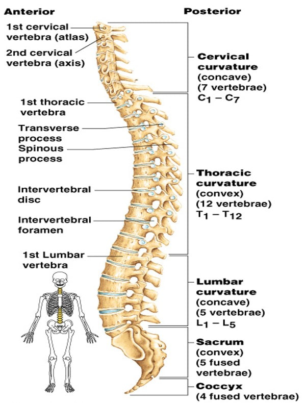
Vertebral Column
The vertebral column usually consists of 33 vertebrae: 24 presacral vertebrae (7 cervical, 12 thoracic, and 5 lumbar) followed by the sacrum (5 fused sacral vertebrae) and the coccyx (4 frequently fused coccygeal vertebrae).
The vertebral column usually consists of 33 vertebrae: 24 presacral vertebrae (7 cervical, 12 thoracic, and 5 lumbar) followed by the sacrum (5 fused sacral vertebrae) and the coccyx (4 frequently fused coccygeal vertebrae).

Atlas (C1)
|

Axis (C2)
|

|

|

Bony Thorax
The bony thorax is formed by the sternum, 12 pairs of ribs, and 12 thoracic vertebrae.
The bony thorax is formed by the sternum, 12 pairs of ribs, and 12 thoracic vertebrae.

Appendicular Skeleton Part III
Appendicular Skeleton
The appendicular skeleton is divided into six major regions: Shoulder girdles (4 bones) - Left and right clavicle (2) and scapula (2). Arms and forearms (6 bones) - Left and right humerus (2) (arm), ulna (2) and radius (2) (forearm).
The appendicular skeleton is divided into six major regions: Shoulder girdles (4 bones) - Left and right clavicle (2) and scapula (2). Arms and forearms (6 bones) - Left and right humerus (2) (arm), ulna (2) and radius (2) (forearm).


|

|
Upper Appendicular Bone Markings


|
|
|
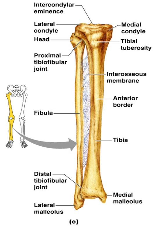
Lower Appendicular Skeleton
The bones of the pelvis connect the bones of the lower limbs to the axial skeleton.
The bones of the pelvis connect the bones of the lower limbs to the axial skeleton.

|

|

|
|

